
A family history of osteoporosis or fragility fracture can make an earlier bone-health baseline valuable. This guide explains when women and men may consider monitoring, which additional risks matter, why REMS offers advantages for proactive assessment, and how to plan meaningful follow-up.
The Quick Answer by Age and Risk Level
If you are wondering at what age to start bone monitoring with family history, the answer depends on life stage, family history, and the presence of additional risks. A younger adult with no known risk factors may use an assessment to document peak bone mass, while someone with a parental hip fracture, early menopause, long-term steroid use, or a previous fragility fracture may need closer attention much sooner.
Women should establish a bone-health baseline by perimenopause, with earlier assessment when family history or other meaningful risk factors are present. Men should receive their first assessment during their 50s, or sooner when their risk profile warrants it.
This prevention-first approach makes future results more meaningful because changes can be compared with a personal baseline rather than interpreted as a single isolated measurement.
At What Age to Start Bone Monitoring With Family History?
Bone monitoring should begin early enough to create a useful baseline, not only after age-based routine screening begins. For women, the transition into perimenopause is an important time to establish that baseline because bone loss can accelerate around the menopausal transition. Women with a family history of osteoporosis, a previous fracture, early menopause, or another meaningful risk factor may benefit from being assessed sooner.
Men should establish their first bone-health baseline during their 50s, with earlier assessment when family history or other significant risk factors are present.
There is also value in understanding bone health much earlier. Bone mass generally reaches its highest level around age 30. An assessment around this stage of adulthood can establish a personal reference point before age-related bone loss becomes more significant.
Family history makes early monitoring especially useful. A parent or sibling with osteoporosis or a fragility fracture may indicate that waiting for a conventional age threshold could mean missing an opportunity to identify changes sooner.
For an age-by-risk breakdown, when you should get a bone density test offers a clear, evidence-based way to understand the right timing.
| Age or life stage | Family history | Added risk factors | Suggested next step |
| Around age 30 | Any | Especially useful when risk factors or proactive monitoring goals are present | Consider documenting an early-adult bone-health baseline |
| 30s or 40s | Yes | One or more | Discuss an earlier baseline assessment |
| Perimenopause | Any | Any | Consider beginning regular bone monitoring |
| Postmenopause | Any | Any | Continue monitoring based on findings and individual risk |
| Men in their 50s | Any | Any | Consider a first bone-health assessment |
| Any age | Yes | Previous fracture, long-term steroid use, early menopause, low body weight, or another significant risk | Arrange an individualized assessment sooner |
Why Family History Changes the Starting Point
Family history is more than background information. A 2025 meta-analysis involving 350,542 men and women from 42 international cohorts found that parental hip-fracture history was associated with a higher risk of future hip fracture. Bone mineral density and fracture risk are shaped by genetic, medical, environmental, and lifestyle factors.
Research has identified a significant association between the bone mineral density of parents and their adult children. Family history does not determine what will happen, but it can strengthen the case for establishing a baseline earlier, especially when it appears alongside other recognized risks.
Bone mineral density and fracture risk are shaped by genetic, medical, environmental, and lifestyle factors. Family history does not determine what will happen, but it can strengthen the case for establishing a baseline earlier, especially when it appears alongside other recognized risks.
Family history becomes more informative when the details are specific. A parent or sibling who experienced a hip fracture after a minor fall provides more useful risk information than a general statement that a distant relative had “weak bones.”
Before speaking with a clinician or bone-health assessment provider, try to identify:
- Which parent, sibling, or other close relative had osteoporosis or low bone density
- Whether the person experienced a hip, wrist, spine, or shoulder fracture
- Approximately how old the person was when the fracture occurred
- Whether the fracture followed a minor fall or routine movement
- Whether several close relatives experienced similar fractures
- Whether early menopause, low body weight, or prolonged medication use also occurred in the family
These details do not predict your outcome on their own. They help create a clearer personal risk picture and may explain why an earlier REMS baseline is worth considering rather than waiting for a general age threshold.
What Age Should I Start If Osteoporosis Runs in My Family?
There is no single starting age that applies to everyone with a family history of osteoporosis. The most useful approach is to establish a baseline before substantial bone loss is likely to occur.
For women, bone monitoring should begin by perimenopause rather than being delayed until later adulthood. An earlier baseline is appropriate when family history appears alongside early menopause, low body weight, prolonged steroid use, a previous low-impact fracture, or another recognized risk.
Men should establish an initial baseline during their 50s, with earlier monitoring when family history or additional risks are present. Adults around age 30 may also choose to document their peak bone mass, creating a valuable reference point for future monitoring.
The purpose of an early assessment is not to assume that bone loss is already present. It is to understand your starting point and make future changes easier to identify.
Why a Baseline Around Age 30 Can Be Valuable
The timing of peak bone mass varies by sex and skeletal site. Most bone mass is acquired during adolescence, but smaller gains can continue through the 20s, with some skeletal sites reaching peak levels around age 30. This makes early adulthood a valuable time to document a personal bone-health baseline before later hormonal and age-related changes become more influential.
A baseline at this stage may be particularly useful for someone with a strong family history, a history of restrictive eating, low body weight, repeated stress fractures, early hormonal disruption, long-term medication use, or another factor that may affect bone development.
Not everyone requires frequent assessments beginning at age 30. The value lies in having a personal reference point that can support more meaningful comparisons during perimenopause, the 50s, or any later period of increased risk.
What Family History and Risk Factors Actually Matter?
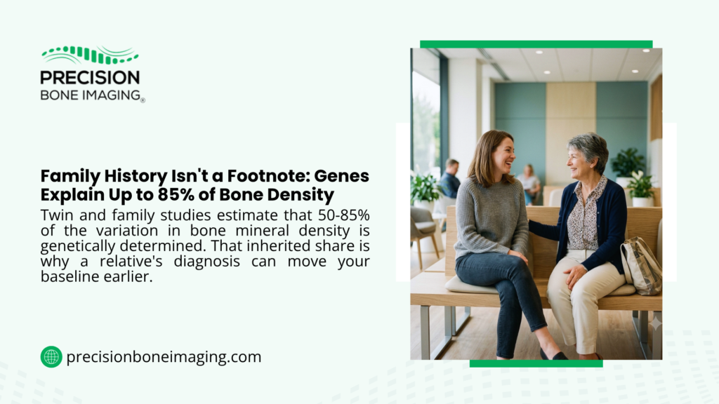
Not all family history carries the same clinical weight. Knowing a parent fractured a hip at 62 carries more weight than a vague sense that “bone problems run in the family.” For a deeper look at how hereditary factors influence bone density, whether osteoporosis is genetic focuses on the genetic side of this risk in more detail.
| Risk profile | Example | Why it matters |
| Family history alone | A parent diagnosed with osteoporosis | Provides a reason to discuss an earlier baseline |
| Family history plus one additional risk | A parent with hip fracture plus low body weight | Strengthens the case for earlier assessment |
| Family history plus several risks | Multiple affected relatives, previous fracture and prolonged corticosteroid use | Supports prompt, individualized evaluation |
| Previous adult fragility fracture | A wrist fracture after a minor fall | May indicate reduced skeletal strength even before other symptoms appear |
| Hormonal risk | Early menopause, menstrual disruption or low testosterone | Hormonal changes can affect bone remodeling |
| Lifestyle or nutritional risk | Smoking, heavy alcohol consumption, restricted eating or physical inactivity | May add modifiable risks to inherited susceptibility |
Additional Risks That Can Move the Timeline Earlier
Recognized factors associated with osteoporosis or fracture risk include:
- Low body weight or a small body frame
- Early menopause or low estrogen levels
- Low testosterone in men
- A previous fracture from minor trauma
- Prolonged use of glucocorticoid medications
- Smoking
- Long-term heavy alcohol consumption
- Physical inactivity or prolonged bed rest
- Low calcium, vitamin D or protein intake
- Eating disorders or excessive dietary restriction
- Certain endocrine, gastrointestinal, inflammatory or kidney conditions
- Medical treatments that can affect hormones or bone metabolism
These factors should be considered together rather than treated as isolated checklist items.
Why a Personal Baseline Can Be More Useful Than an Age Threshold
Age-based recommendations are designed for population-level screening. A personal monitoring plan asks a different question: when would an individual measurement become useful for understanding future change?
A baseline collected before an expected period of bone loss may help answer:
- Were bone-density concerns already present in early adulthood?
- Did measurements change during perimenopause or menopause?
- Are lifestyle and nutritional strategies associated with a measurable trend?
- Does one anatomical site show more concern than another?
- Does the Fragility Score add information about skeletal fragility beyond the density result?
What Type of Bone Monitoring Should You Consider?
REMS vs DEXA: Which Provides a More Complete Proactive Assessment?
DEXA uses low-dose X-rays to measure bone mineral density, while REMS provides a radiation-free assessment of bone mineral density and skeletal fragility at the lumbar spine and proximal femur. It reports bone mineral density and provides a Fragility Score related to skeletal fragility and characteristics associated with bone quality.
| Feature | REMS | DEXA |
| Technology | Ultrasound-based radiofrequency analysis | Low-dose X-ray imaging |
| Ionizing radiation | None | Yes, at a low dose |
| Bone mineral density | Measured at the lumbar spine and proximal femur | Measured at the lumbar spine and hip |
| Additional skeletal-fragility information | Provides a Fragility Score related to skeletal fragility and characteristics associated with bone quality | Standard DEXA does not provide the REMS Fragility Score |
| Use for proactive follow-up | Well suited to repeat assessment because it is radiation-free | Repeat timing is commonly determined by clinical need and conventional monitoring protocols |
| Portability | Portable and suitable for partner clinics or mobile scan events | Usually performed using fixed equipment in an imaging facility |
| Potential artifact management | REMS algorithms are designed to identify and exclude signals associated with some artifacts and degenerative changes | Degenerative changes, positioning, and other artifacts can sometimes affect interpretation |
| Results through Precision Bone Imaging | Immediate, color-coded report with an explanation of key findings | Reporting procedures vary by imaging provider |
For proactive monitoring, REMS offers several clear advantages: it does not use ionizing radiation, reports bone mineral density, adds a Fragility Score related to skeletal fragility, and can be repeated when clinically or personally useful.
Research has found that REMS can provide fracture-risk information and has demonstrated strong repeatability in studied populations. However, REMS and DEXA are different technologies, and their measurements should not be treated as directly interchangeable in every person or clinical situation.
Bone Density and Skeletal Fragility in Plain English
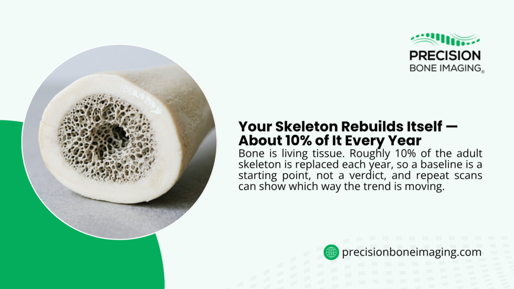
Bone mineral density describes how much mineral is measured within a defined area of bone. Bone strength, however, is influenced by more than density alone. Bone structure, turnover, accumulated damage, and other characteristics commonly discussed as aspects of bone quality can also contribute to skeletal fragility.
REMS supplements its density measurement with the Fragility Score, a parameter developed to reflect spectral characteristics associated with bone microarchitecture and skeletal fragility. Research suggests that the score may add information about fracture susceptibility, including in some cases where density alone does not provide the complete risk picture.
This means two people with similar density findings may not necessarily have identical skeletal strength or fracture risk. A more complete assessment considers density, quality, age, fracture history, family history, health conditions, and lifestyle together.
Why Does REMS Support Earlier and Ongoing Monitoring?
A proactive baseline is most valuable when it can be compared with future measurements. Because REMS is radiation-free, it removes radiation exposure as a barrier to repeat monitoring. That makes REMS particularly relevant for:
- Women moving through perimenopause or menopause
- Men establishing a baseline during midlife
- Adults with a strong family history
- Athletes monitoring skeletal health
- People making significant nutrition or exercise changes
- People who want to follow bone-health trends over time
- Wellness and longevity clients treating bone health as an important healthspan marker
Whenever possible, follow-up assessments should use the same technology, comparable anatomical sites, and consistent demographic information. This helps reduce avoidable variation and makes trends easier to interpret.
How Often Should Bone Monitoring Be Repeated?
There is no universal follow-up interval that is right for every person. How often you should get a bone density scan depends on initial findings, life stage, fracture history, current risks, and the purpose of monitoring.
| Situation | Potential follow-up approach |
| Early-adult baseline with no immediate concern | Keep the result as a personal reference and determine future timing from changes in health or risk |
| Perimenopause or a changing risk profile | Discuss whether follow-up within approximately 12 months would provide useful trend information |
| Lifestyle or bone-health program in progress | A six- or 12-month REMS assessment may help track measurable change |
| Lower bone mass or elevated Fragility Score | Create an individualized follow-up plan with an appropriate healthcare professional |
| New fracture, rapid height loss or new medical risk | Seek clinical evaluation rather than waiting for a routine re-scan date |
Precision Bone Imaging’s Bone Health Blueprint includes follow-up scans intended to help clients track changes over time. However, a six-month re-scan should not be presented as medically necessary for every person. The interval should have a clear purpose and be interpreted in the context of the individual’s overall health.
What Should You Do After Receiving Your Results?
A bone-health report should lead to an informed plan rather than fear or a rushed decision. The next step depends on the pattern of findings and the individual’s wider risk profile. A useful discussion may cover:
- Resistance training, appropriate impact exercise, and Osteogenic Loading
- Adequate protein and total energy intake
- Individual calcium and vitamin D requirements
- Hormonal or medical factors that could affect bone
- Smoking and alcohol use
- Fall risk, balance, and muscle strength
- Relevant laboratory testing
- Whether referral to a qualified healthcare professional is needed
- An appropriate time for repeat assessment
Disclaimer: This article does not present medication as the automatic response to every result. Decisions about medical treatment should be individualized and discussed with a licensed healthcare professional. Nutrition, exercise, possible underlying causes, personal preferences, and overall fracture risk all belong in that conversation.
What Should You Bring to a Bone-Health Appointment?
| Bring this information | Why it helps |
| Which relatives had osteoporosis or fractures | Clarifies the strength of the family pattern |
| Relatives’ approximate ages at diagnosis or fracture | Helps distinguish earlier fragility from fractures occurring later in life |
| Your previous fractures | A low-impact adult fracture may affect your risk assessment |
| Medication and supplement list | Certain medications and health regimens can influence bone metabolism |
| Menstrual, menopause, or hormone history | Hormonal changes can affect bone remodeling |
| Previous DEXA or REMS reports | Provides context and helps identify possible trends |
| Exercise and nutrition habits | Shows which modifiable factors may deserve attention |
Questions to Ask Your Clinician or REMS Provider
- How does my family history affect the value of establishing a baseline now?
- Which additional risk factors apply to me?
- What do my bone mineral density and Fragility Score show when considered together?
- Do I need further clinical evaluation or laboratory testing?
- Which lifestyle changes are most relevant to my findings?
- When would a follow-up REMS assessment provide meaningful information?
- Should future assessments use the same technology for a clearer comparison?
When Should You Arrange an Assessment Sooner?
Consider arranging an earlier conversation or assessment if family history appears alongside:
- A fracture following a minor fall or routine movement
- Perimenopause, menopause, or early menopause
- Unexplained height loss or a noticeable posture change
- Prolonged corticosteroid use
- Very low body weight or substantial recent weight loss
- An eating disorder or prolonged nutritional restriction
- Menstrual disruption
- Low testosterone
- A condition or treatment associated with bone loss
- Several close relatives with osteoporosis or fragility fractures
Sudden severe back pain, a suspected fracture, or a significant injury requires medical evaluation. A routine bone-health monitoring appointment should not replace urgent or diagnostic medical care.
FAQs
At What Age Should I Start Bone Monitoring With a Family History?
Women should establish a baseline by perimenopause, while those with additional risks should begin earlier. Men should receive their first bone-health assessment during their 50s, or sooner when family history or other concerns increase their risk. An assessment around age 30 can also document an early-adult reference point for future comparison.
Does Having a Parent With Osteoporosis Mean I Need an Assessment Immediately?
Not necessarily, but it is a meaningful reason to review your risk. Timing depends on your age, sex, hormonal stage, previous fractures, body weight, medication exposure, health conditions, and other risk factors.
Should Women Wait Until After Menopause?
No. Perimenopause can be a valuable time to establish a baseline, and women with a family history or additional risks may benefit from assessment before that stage. Hormonal changes associated with menopause can contribute to faster bone loss.
When Should Men With a Family History Begin Monitoring?
Precision Bone Imaging recommends that men establish their first bone-health baseline during their 50s. Men with a family history, previous fracture, low body weight, low testosterone, prolonged corticosteroid use, or another recognized risk should consider beginning earlier.
Why Consider an Assessment Around Age 30?
Peak bone mass is generally established by early adulthood. A REMS assessment around age 30 can document a personal reference point before later hormonal and age-related changes become more influential.
Is REMS Better Suited to Proactive Monitoring Than DEXA?
REMS has important advantages for proactive assessment: it is radiation-free, evaluates bone mineral density at the lumbar spine and proximal femur, and provides a Fragility Score related to skeletal fragility and characteristics associated with bone quality. It is also portable and suitable for repeat monitoring.
DEXA remains widely used in conventional medical pathways, while REMS provides an additional radiation-free option for people who want to monitor bone health proactively.
Can REMS Results Be Compared Directly With DEXA Results?
The two technologies may produce related information, but they are not interchangeable in every person. For clearer trend tracking, use the same assessment technology and follow consistent testing procedures whenever possible.
Take the First Step Toward a Clearer Bone-Health Baseline
A family history of osteoporosis does not guarantee that you will develop the condition or experience a fracture. It does mean that understanding your personal starting point may be more valuable, and may become valuable earlier, than it is for someone without the same background.
Precision Bone Imaging provides comprehensive assessments using FDA-cleared, radiation-free Echolight REMS technology. Each assessment evaluates the lumbar spine and hip region and provides bone mineral density results, a Fragility Score related to skeletal fragility, body-composition information, and an estimate of five-year fracture risk, followed by an immediate, easy-to-understand report.
With scheduled and mobile locations across California, Arizona, Colorado, and a growing partner network, Precision Bone Imaging makes proactive bone-health assessment more accessible.
Book your radiation-free REMS assessment to establish a baseline, understand your bone mineral density and Fragility Score, and make your next bone-health decisions with greater clarity.
Note: This information is educational and does not replace diagnosis, treatment, or individualized advice from a licensed healthcare professional.